

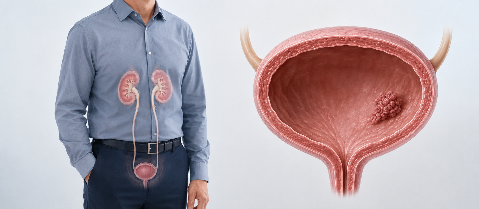
Bladder cancer is a malignant tumor that develops from the cells lining the inside of the bladder. Its most common symptom is painless, visible blood in the urine. A definitive diagnosis is usually made after cystoscopy and transurethral resection of the bladder tumor (TURBT).
The most important distinction for treatment is whether the tumor has invaded the bladder muscle. Non-muscle-invasive disease is generally managed with bladder-preserving endoscopic and intravesical therapy, while muscle-invasive or very high-risk disease may require radical cystectomy, lymph node dissection, and urinary diversion.
Last medical review: 2026
Symptoms of bladder cancer
Blood in the urine — pink, red, or dark discoloration, often painless and sometimes intermittent — is the most common symptom and should always be investigated, even if it resolves on its own. Other symptoms can include frequent urination, urgency, burning on urination, or a weak stream. Advanced disease may cause flank pain, bone pain, leg swelling, poor appetite, or unexplained weight loss.
Risk factors
Smoking is the most important preventable risk factor, since carcinogens in tobacco smoke are excreted in urine and contact the bladder lining. Occupational exposure to certain dyes, rubber, leather, textile, or industrial chemicals raises risk, as does prior pelvic radiotherapy or certain drugs such as cyclophosphamide. Chronic bladder irritation, long-term catheter use, and a personal history of urothelial cancer also increase risk. Absence of a risk factor does not exclude bladder cancer.
How is bladder cancer diagnosed?
Evaluation includes clinical assessment, urine tests including cytology, and imaging — typically contrast CT urography to assess the upper urinary tract and bladder. Cystoscopy allows direct visualization of the bladder lining, and TURBT is both diagnostic and therapeutic: the visible tumor is removed endoscopically, and the specimen — including muscle tissue, to reliably assess for muscle invasion — is sent for pathology. In select patients (high grade, T1, incomplete initial resection, or no muscle in the specimen), a second TURBT may be recommended.
What do stage and grade mean?
Ta tumors are confined to the bladder lining; carcinoma in situ (CIS) is a flat, high-grade lesion with a meaningful risk of progression; T1 has invaded the connective tissue beneath the lining but not the muscle; T2 has invaded the muscle layer; T3 extends into the surrounding fat; and T4 involves adjacent organs. Grade describes how aggressive the cells look under the microscope, while stage describes how far the tumor has spread — both are needed to plan treatment.
Treatment of non-muscle-invasive bladder cancer
The goal is complete removal of visible tumor, reducing recurrence, and preventing progression to muscle-invasive disease. Depending on risk group after TURBT, options include a single dose of intravesical chemotherapy shortly after resection, surveillance cystoscopy for low-risk tumors, a course of intravesical chemotherapy for intermediate-risk disease, and intravesical BCG for high-risk tumors, T1 high-grade, or CIS. Very high-risk disease not responding adequately to BCG may be considered for early radical cystectomy.
Treatment of muscle-invasive bladder cancer
For muscle-invasive disease without distant metastasis, the main curative options are radical cystectomy with pelvic lymph node dissection and urinary diversion, generally combined with perioperative systemic therapy for eligible patients, or, in carefully selected cases, bladder-preserving trimodal therapy combining maximal TURBT with concurrent chemotherapy and radiotherapy.
Robotic radical cystectomy
Robotic radical cystectomy removes the bladder and regional lymph nodes through small incisions, with the surgeon controlling every instrument movement from a console. Potential advantages include magnified three-dimensional visualization, precise dissection in the deep pelvis, and less blood loss than open surgery; current comparative data show broadly similar 90-day complication rates, surgical margins, and mid-term cancer outcomes between robotic and open approaches. Surgeon and center experience with cystectomy and diversion remain the most important factors in outcome.
Urinary diversion after cystectomy
Once the bladder is removed, a new pathway for urine is created. An ileal conduit uses a short segment of small bowel to carry urine to a stoma on the abdominal wall, draining continuously into an external bag — a well-established option offering predictable drainage and standardized care. An orthotopic neobladder creates an internal reservoir from bowel that is connected to the urethra, allowing voiding without an external bag, but it requires suitable urethral, kidney, and cognitive function, timed voiding training, and sometimes intermittent catheterization; not every patient is a candidate. The choice depends on cancer safety first, then anatomy, organ function, and the patient's priorities and ability to manage the care each option requires.
Follow-up after treatment
Non-muscle-invasive disease is followed with regular cystoscopy, urine cytology, and upper tract imaging based on risk. After radical cystectomy, follow-up includes cancer surveillance imaging, kidney function monitoring, evaluation of the diversion, and attention to metabolic changes and quality of life, including continence and sexual function.
Frequently asked questions
Does blood in the urine always mean bladder cancer?
No, but it should always be investigated, including when it occurs only once and resolves on its own. Causes range from infection and stones to bladder cancer, so evaluation with imaging and cystoscopy is important.
Does the whole bladder always need to be removed?
No. Most non-muscle-invasive tumors are managed with TURBT and intravesical therapy. Radical cystectomy is generally reserved for muscle-invasive disease or very high-risk tumors that do not respond to bladder-preserving treatment.
Is life without a bladder possible?
Yes. After cystectomy, urine is diverted through an ileal conduit or a neobladder. Both require an adjustment period and long-term follow-up, and the right option depends on the patient's anatomy and priorities.
Source: European Association of Urology (EAU) Guidelines on Non-muscle-invasive and Muscle-invasive Bladder Cancer, 2026. This content is for general information only and does not replace individualized medical evaluation. Prepared and medically reviewed by Professor Cenk Acar, Urologic Oncologist.