

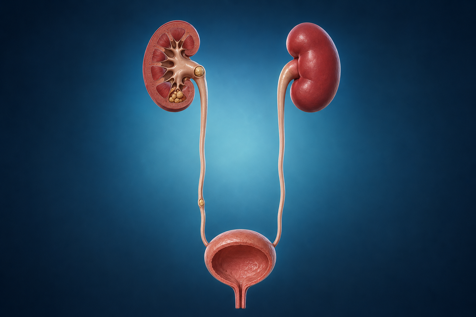
A kidney stone forms when certain minerals in the urine crystallize into a hard mass within the kidney or urinary tract. Some small, non-obstructing stones can be monitored or pass on their own; others causing pain, obstruction, infection, or declining kidney function require treatment.
Treatment is not chosen by stone size alone — its location, number, density on CT, urinary tract anatomy, associated infection, kidney function, medications, and the patient's priorities are all considered together.
Last medical review: 2026
What causes kidney stones?
Stone disease is rarely due to a single cause. Genetic predisposition, low fluid intake, dietary habits, certain metabolic or bowel conditions, and urinary tract anatomy can all contribute. Low urine volume — from inadequate fluid intake, heat exposure, intense exercise, or heavy sweating — concentrates the urine; a family history or stone onset in childhood or young adulthood warrants closer evaluation; and high salt or, in some people, high animal protein intake, obesity, and gout can raise risk. Certain bowel diseases and surgeries can increase oxalate absorption, and some medications and supplements can promote stone formation in susceptible individuals.
Types of kidney stones
Calcium oxalate/phosphate stones are the most common group, influenced by urinary calcium, oxalate, citrate, and sodium levels. Uric acid stones are linked to acidic urine, gout, obesity, or metabolic syndrome, and some can be dissolved with medication. Infection stones are associated with certain urease-producing bacteria, can grow quickly, and may fill the kidney's collecting system. Cystine stones are rare, arise from an inherited condition called cystinuria, and tend to recur. Stone appearance alone does not reliably indicate its type — laboratory analysis of a passed or removed stone strengthens prevention planning.
Symptoms of kidney stones
A small stone sitting quietly in the kidney may cause no symptoms. When a stone moves into the ureter and obstructs urine flow, pain typically begins suddenly, waxes and wanes in waves, and makes it difficult to find a comfortable position. It commonly starts below the ribs and radiates to the lower abdomen, groin, or genitals. Other symptoms can include visible or microscopic blood in the urine, nausea and vomiting, urinary urgency and frequency as the stone nears the bladder, and fever or chills suggesting an associated infection — which, together with obstruction, is a urologic emergency requiring prompt evaluation.
How are kidney stones diagnosed?
Diagnosis combines history, examination, urine and blood tests, and imaging. Ultrasound involves no radiation and is often the first imaging choice, particularly in pregnancy and children. Low-dose non-contrast CT provides detailed information on stone location, size, and density, and is often used for acute flank pain when it will influence the treatment plan. Imaging is chosen to balance diagnostic accuracy against cumulative radiation exposure, especially in young or recurrent stone formers.
Does every kidney stone need treatment?
No. Small, non-obstructing, asymptomatic stones can often be monitored with periodic imaging. Growth, pain, bleeding, obstruction, infection, threatened kidney function, or significant impact on work or travel are reasons to consider active treatment. For ureteral stones, the chance of spontaneous passage increases as the stone is smaller and closer to the bladder, but size alone cannot guarantee a timeframe — adequate pain control, absence of infection, and preserved kidney function are the key requirements for continued observation. In selected lower ureteral stones of 5–10 mm, alpha-blocker medication may be prescribed to help facilitate passage, though it is not appropriate for everyone.
Treatment options
Observation or medical expulsive therapy suits small, uncomplicated ureteral stones likely to pass, or select asymptomatic kidney stones, with regular follow-up. Shock wave lithotripsy (SWL) focuses shock waves from outside the body to fragment suitable stones without any instrument entering the body, though multiple sessions may be needed and success decreases with harder, larger, or lower-pole stones. Ureteroscopy (URS) with laser passes a scope through the urinary tract to fragment or remove ureteral stones, generally with a higher single-session clearance rate than SWL. Flexible ureteroscopy (RIRS) reaches stones within the kidney, typically up to about 2 cm, using a flexible scope and laser fragmentation. PCNL/mini-PCNL creates a small tract through the skin into the kidney and is generally the first choice for stones larger than 2 cm or those filling the collecting system, offering high clearance rates but with a somewhat higher risk of bleeding and infection than the endoscopic retrograde options.
Preventing recurrence
Removing a stone treats the current problem but does not necessarily remove the underlying tendency to form stones, so stone analysis and basic blood and urine testing are valuable even after a first stone. Patients with recurrent or bilateral stones, onset at a young age, a family history, a solitary kidney, uric acid, cystine, or infection stones, or an underlying bowel or kidney condition are considered higher risk and may benefit from detailed metabolic evaluation, typically with 24-hour urine collections, to guide targeted prevention with adequate hydration, dietary adjustment, and, when indicated, medication such as potassium citrate or a thiazide diuretic.
General prevention measures
Adequate fluid intake spread through the day — enough to produce pale urine, individualized for heart and kidney conditions — is the single most consistent recommendation. Reducing excess sodium, maintaining normal dietary calcium rather than restricting it unnecessarily, moderating animal protein according to stone type, and maintaining a healthy weight and activity level all support prevention, alongside periodic follow-up guided by stone analysis and metabolic results.
Frequently asked questions
Does every kidney stone cause pain?
No. A stone that stays within the kidney without obstructing urine flow can be completely silent and may only be found incidentally on imaging done for another reason.
Will a small stone pass on its own?
Smaller stones, particularly those closer to the bladder, are more likely to pass spontaneously, but this cannot be guaranteed by size alone. Adequate pain control, absence of infection, and preserved kidney function are needed to safely continue observation.
Is SWL or laser treatment better?
Neither is universally better. SWL avoids inserting instruments into the body but may need repeat sessions, while endoscopic laser treatment generally offers a higher chance of stone-free status in a single procedure. The right choice depends on stone size, location, density, and patient factors.
Do kidney stones come back after treatment?
Removing a stone does not remove the tendency to form new ones. Stone analysis, metabolic evaluation in higher-risk patients, and consistent prevention measures reduce — but cannot completely eliminate — the risk of recurrence.
Source: European Association of Urology (EAU) Guidelines on Urolithiasis, 2026. This content is for general information only and does not replace individualized medical evaluation. Prepared and medically reviewed by Professor Cenk Acar, Urologist.