

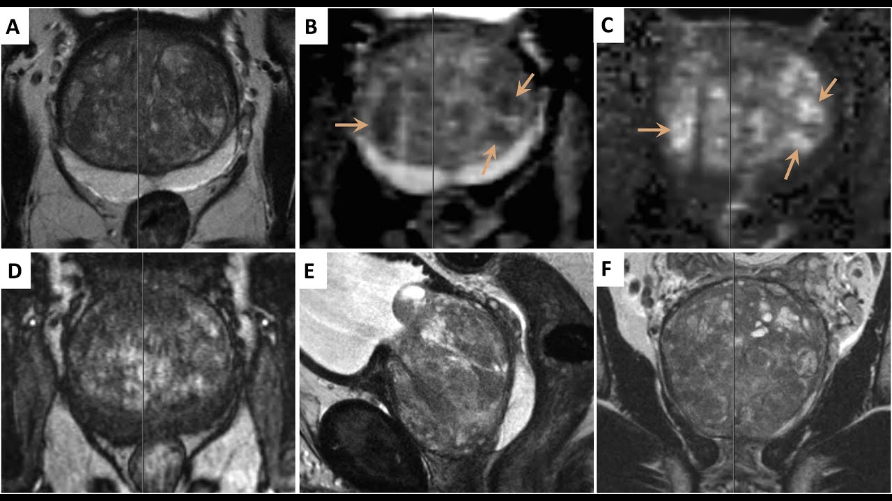
MRI fusion prostate biopsy is a technique in which suspicious areas identified on multiparametric prostate MRI are precisely merged with real-time transrectal or transperineal ultrasound imaging, allowing targeted sampling of the exact area of concern.
In our practice, the procedure is performed using the transperineal approach — without the needle passing through the rectum — under general anesthesia or sedation.
Last medical review: 2026
What is MRI fusion prostate biopsy?
It is a combined diagnostic technique that merges multiparametric MRI data with ultrasound imaging. Dedicated software fuses the MRI images with real-time ultrasound, allowing tissue to be sampled precisely from the suspicious area rather than blindly.
Who may be offered this procedure?
It is generally offered to patients with elevated PSA and/or suspicious findings on prostate MRI — typically a PI-RADS score of 3 or higher — and in cases where a prior standard biopsy did not yield a result consistent with the clinical picture.
What does the PI-RADS score mean?
PI-RADS (Prostate Imaging – Reporting and Data System) is a standardized 1-to-5 scale reflecting the likelihood of clinically significant prostate cancer based on MRI findings. A higher score generally increases suspicion and strengthens the case for a targeted biopsy, though the decision also depends on PSA, prostate volume, and other clinical factors.
Why is the transperineal approach used?
With the transperineal approach, the needle passes through the perineal skin rather than through the rectum. This reduces the risk of infectious complications and allows more precise, even coverage of the entire gland, including the anterior zone, which is harder to reach transrectally.
What is the difference between targeted and systematic biopsy?
Targeted (fusion) biopsy samples tissue directly from the area flagged as suspicious on MRI. Systematic biopsy takes samples according to a standard pattern across different regions of the prostate, independent of MRI findings. The two are typically combined, which improves diagnostic accuracy and reduces the chance of missing a significant tumor.
How is the procedure performed?
After anesthesia or sedation is administered, the physician uses an ultrasound probe together with the fused MRI images to precisely sample the suspicious area or areas, along with systematic samples from the rest of the gland when appropriate. The procedure usually takes 20–40 minutes, though preparation, anesthesia, and post-procedure observation add to the total time.
What happens after the biopsy?
Most patients go home the same day or the following day. Mild discomfort, and a small amount of blood in the urine or semen for a few days, are expected and usually resolve on their own. Fever, severe pain, difficulty urinating, or heavy bleeding should prompt immediate contact with the treating physician.
How is the result assessed?
The tissue obtained is examined by a pathologist, who determines the presence, extent, and aggressiveness of any tumor using the Gleason score and ISUP grade group. Results are typically available within 1–2 weeks and discussed at an in-person consultation. A negative result substantially lowers the likelihood of clinically significant cancer but does not exclude it completely — if clinical suspicion remains, active surveillance or a repeat biopsy may be recommended.
Frequently asked questions
Is MRI fusion biopsy painful?
The procedure is performed under general anesthesia or sedation, so discomfort during the procedure itself is minimal. Mild soreness or urinary symptoms for a few days afterward are common and expected.
How long is recovery?
Most men return to routine activity within a day or two, though heavy exertion is usually avoided for a short period as advised by the treating physician.
Is hospital admission required?
Most patients are discharged the same day or the next day; overnight observation depends on the anesthesia used and individual factors.
This content is for general information only and does not replace individualized medical evaluation. Prepared and medically reviewed by Professor Cenk Acar, Urologic Oncologist.